Avian musculature contains an increased density of muscle fibres and less connective tissue than that of mammals.
 |
 |
|
|
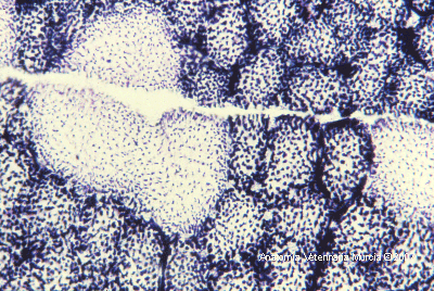
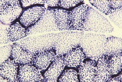
Transverse histological section of muscle fibres
stained for NADH-TR. The mitochondria appear stained blue. The connective
tissue does not stain (white). White glycolitic fibres, dark oxidative
fibres. |
Intramuscular fat is scarce and the colour of the muscle is dependent on the region of the body and the species. In flying birds the pectoral musculature is redish in colour, indicating a large number of muscle fibres rich in myoglobin (oxidative aerobic metabolism),
whereas in birds which do not have the ability to fly, this musculature is pale due to the predominance of white glycolytic muscle fibres (anaerobic metabolism). The main muscles involved in flight are the pectoral muscles (breast). There is a superficial pectoral muscle and a deep pectoral or supracoracoid muscle. The superficial pectoral muscle extends from the keel of the sternum, the clavicle and the sternocoracoclavicular membrane, to the pectoral crest of the humerus. This is the main depressor of the wing.
The deep pectoral muscle lifts the wing from the keel of the sternum. It develops a tendon that passes through the triosseal canal to the dorsal surface of the humerus.
This tendon can be ruptured if a traumatic
event occurs (flying into a window, for example) and the classic presentation
is seen as the bird being unable to lift its wing. In this case repair of
the tendon is required. However, by bandaging the affected wing the usual
formation of a callus in the trioseum canal results in the same outcome.
The pectoral musculature should always be examined as it indicates the nutritional
state of the bird. It can also be used as a site for intramuscular injections.
The caudal part of the superficial pectoral muscle is recommended as the cranial
part has an abundant blood supply, and this increases the risk of injecting
substances directly into the bloodstream.
Among the muscles of the wing, the extensor
carpi radialis should be emphasized. It originates from the medial epicondyle
of the humerus, extends over the cranial surface of the carpal joint and ends
in the carpometacarpal extensor apophysis.
Flight is also facilitated by the propatagium,a cutaneous triangular fold in the cranial part of the wing, which extends between the the shoulder and carpus.
On the ventral surface of the propatagium access to the cutaneous ulnar vein is possible, making it an appropriate site for venipuncture over the site of the elbow joint.
The main function of the muscles in the
pelvic limb is to maintain the body upright and in equilibrium, as well as
being of use during locomotion. The pelvic muscles can be used for the administration
of intramuscular injections, although due to a portal-renal venous system
any drug administered here travels via the kidney before reaching the systemic
circulation.
Most perch and prey birds have a reciprocal apparatus which causes the flexion
of interphalangial joints when the tarsal joint is flexed. As a result of
this the digital flexor tendon passively clamps the digits around the perch
when the tarsus is flexed. This mechanism should be remembered when attempting
to undo the grasp of a large bird.
On the medial aspect of the tarsal joint of large birds the caudal tibial
vein can be accessed for venipuncture. Also, for diagnostic purposes, for
example Marek’s disease, it is useful to locate the sciatic nerve at
its passage through the thigh, caudal to the femur, covered by the musculature
of the region.
| |
|
Finally, ossification of the flexor tendons of the digits (gastrocnemius m.,
superficial and deep digital flexor mm.) is a sign of ageing found in some
birds.
The trunk muscles are less important but those of the neck are highly developed
due to the mobility of this region. The abdominal and intercostal muscles
are reduced to thin layers.