Instructions
In order to
use the program and achieve the maximum benefit, the following should
be considered:
The work is based on an anatomical image which is accompanied by a glossary
of terms, "Anatomical Structures", situated at the right hand
side of the image.
A). – Anatomical image. As shown in the example, running the cursor
over the image will cause the structure to become highlighted (coloured)
and at the right hand side of the screen the name of the structure upon
which the cursor is positioned will appear. In this way the dissection
plans can be studied interactively. For example, in Image 1 the cursor
(a black arrow), is placed on an area of the heart, which appears highlighted.
Immediately the name of the structure appears to the right of the screen
(glossary of terms), in this case the "left ventricle and epicardium".
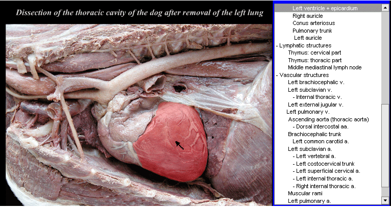
Image 1
B). – Anatomical Structures. As is shown in another example, using
the cursor to click on the name of the structure of interest from the
glossary of terms (to the right of the screen) will cause the relevant
structure to appear highlighted (coloured). For example, in images 2 and
3. By clicking with the black arrow on "left ventricle and epicardium",
the text will remain highlighted and on the image the corresponding structure
will appear coloured. This allows a way of testing oneself, as until the
name in the glossary is clicked on the structure will not become highlighted
(before clicking attempt to mentally identify the structure)
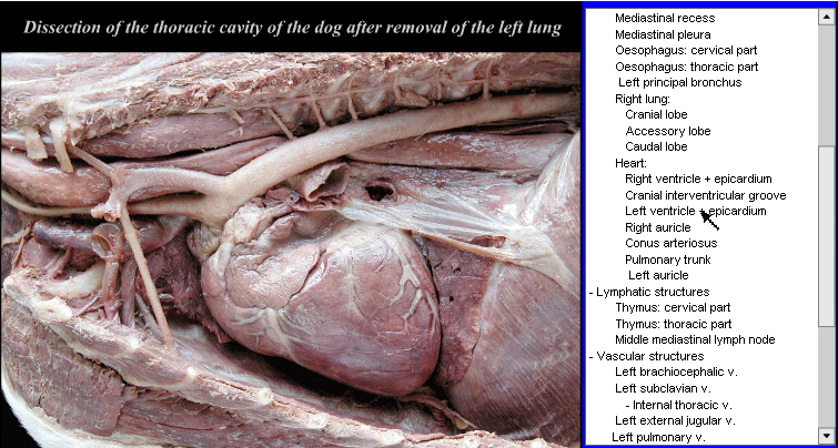
Image 2
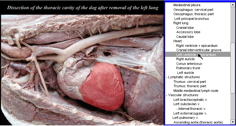
Image 3